Training for complex procedures - By enabling surgeons to perform the procedure in a controlled, physical environment, these models improved understanding, confidence, and skill development - providing a more effective alternative to purely observational or theoretical training.
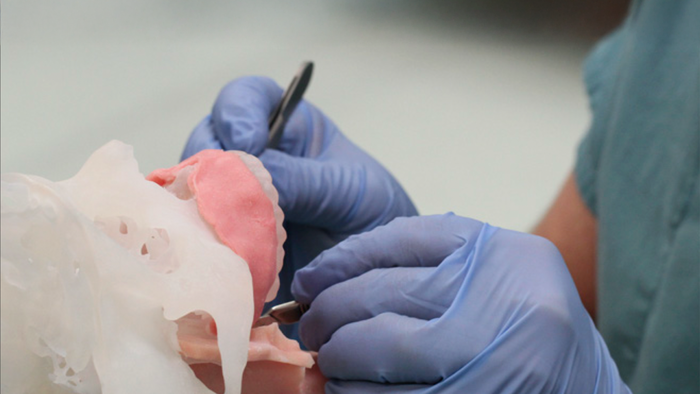
Hands-on testing with the surgeons was essential to bridging the gap between technical design and clinical practice. We used these prototyping sessions to observe how the models were handled, where assumptions broke down, and how the procedure was interpreted in real time.
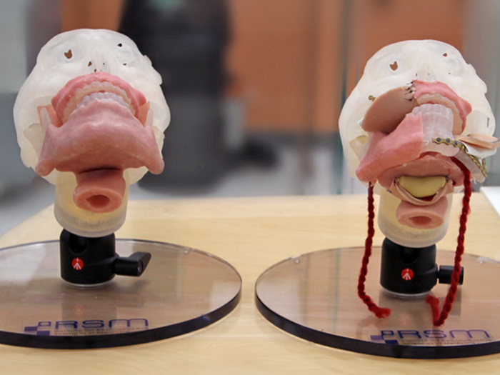
Before and after the initial surgical simulation test run
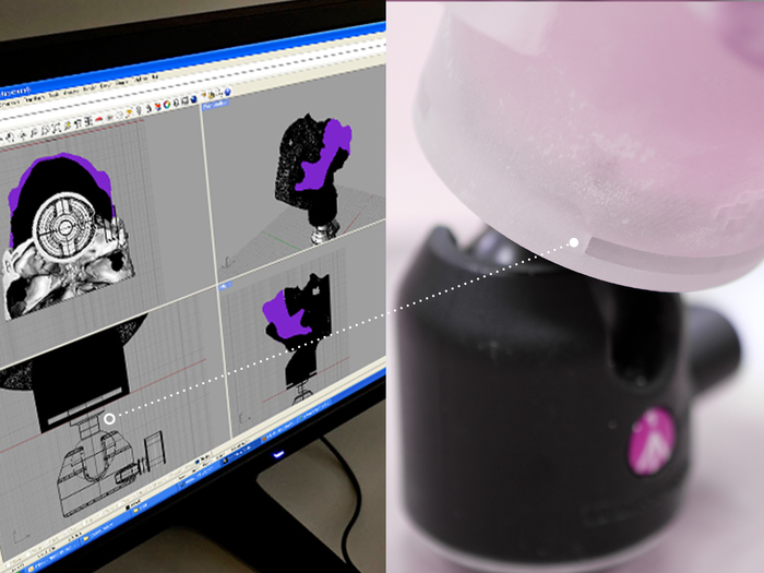
I designed the skull to connect to a camera mount, allowing it to pivot in all directions for easy handling and demonstration during training. Designed for usability and education, the jaw features a removable ball joint, while the base uses a sliding assembly to keep the skull secure yet easy to assemble and disassemble.
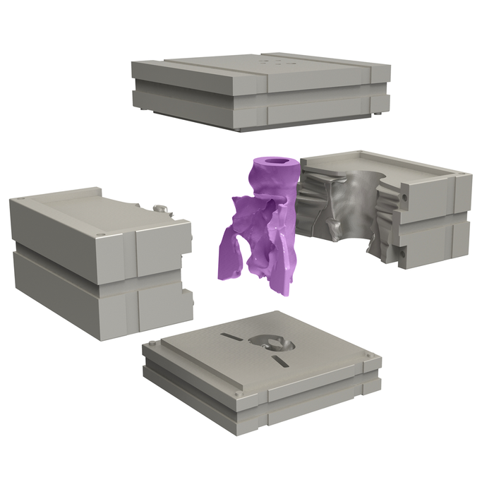
Designing the molds was an adventure. Complex anatomy leads to challenges in releasing the molds.
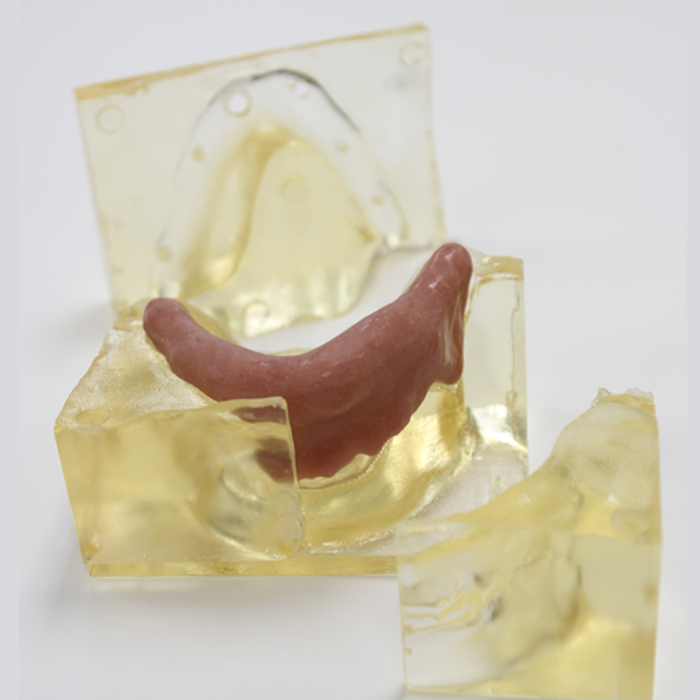
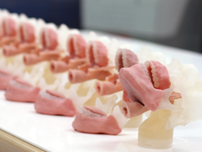
In-house resin 3D prints were used in the mold making process for rapid iterations.
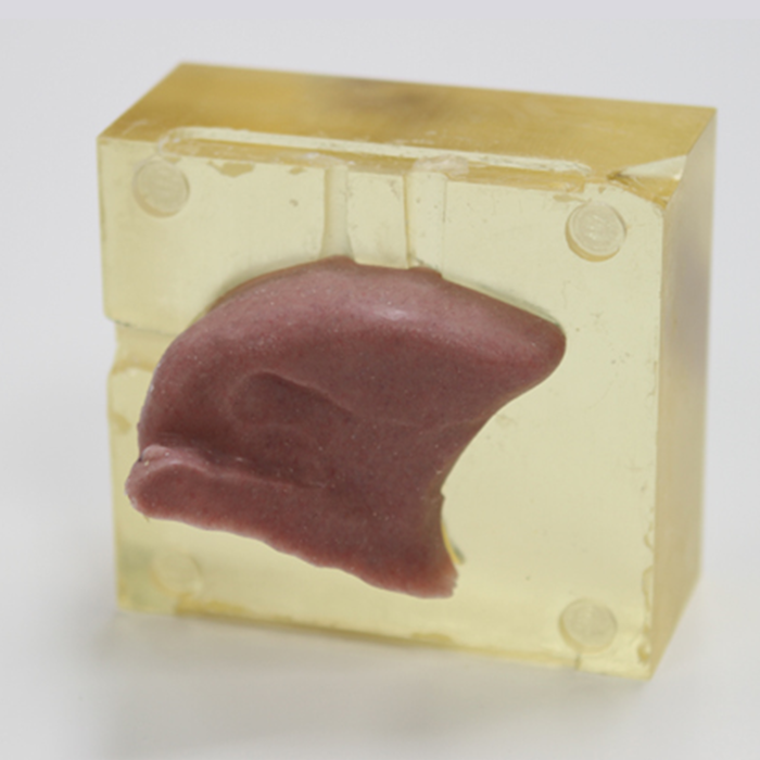
Different parts, like the tongue, required different feel and softness to simulate actual anatomy.
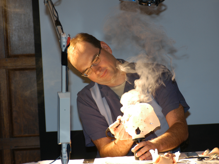
The surgical training seminar combined an overhead camera system with individual workstations, allowing each participant to follow along and practice in real time. Dr. Harris guided the procedure throughout the course, demonstrating techniques, answering questions, and assessing proficiency as surgeons worked through each step.
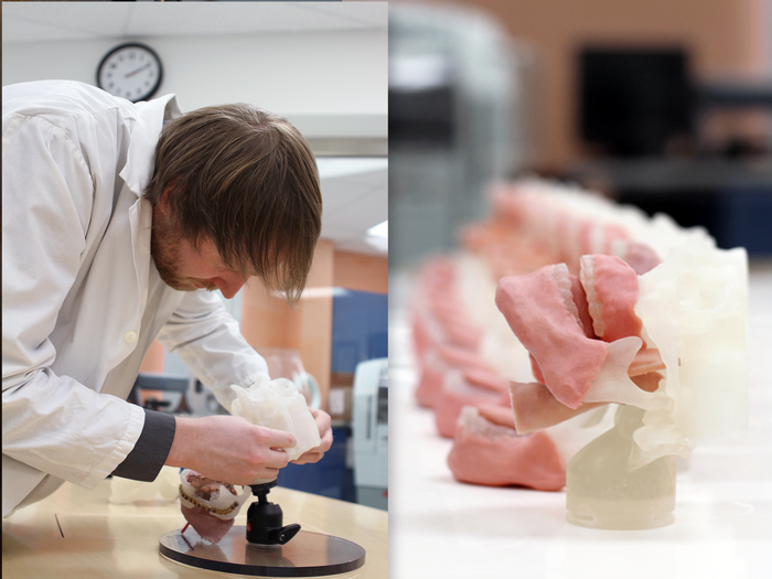
This approach marked a shift from the traditional “see one, do one, teach one” model toward a more structured, simulation-based method focused on repetition, feedback, and measurable skill development.
gLike
Surgical Simulation
Developed in collaboration with Andrew Grosvenor and clinical partners at iRSM, this project focused on creating realistic physical models to support surgical training in complex head and neck reconstruction.
The models were designed to simulate key aspects of the surgery, allowing participants to remove a mock tumor, reconstruct the tongue and the mandible using a fibula graft, and plate the structure as they would in a real procedure.